When scientists mapped all 302 of a nematode worm’s neurons
in 1986 , they’d arrived at a major milestone in brain research. It was the first time a species’ “connectome”—a comprehensive map of neural connections—was documented and analyzed in its entirety.But there was something missing. To understand a species’ nervous system, you need more than just its neural blueprint. You need to know how those neurons work together, stretching from a sensory stimulus all the way to the brain’s interpretation (or, in the case of nematodes, the ganglia’s interpretation). And you need this information in three dimensions, over a large volume, and in real time.
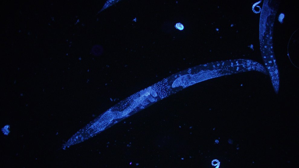
Now, researchers have succeeded in imaging all of C. elegans ‘ neurons in real time. Such 3D “movies” of brains at the millisecond timescale could help scientists understand what type of neural activities result in certain behaviors. It could even help them determine what cells are involved in brain disorders.
Here’s Sara Reardon, writing for Nature:

Vaziri and his colleagues engineered C. elegans so that when a neuron fires and calcium ions pass through its cell membranes, the neuron lights up. To capture those signals, they imaged the whole worm using a technique called light-field deconvolution microscopy, which combines images from a set of tiny lenses and analyses them using an algorithm to give a high-resolution three-dimensional image. The researchers took as many as 50 images per second of the entire worm, enabling them to watch the neurons firing in the brain, ventral cord, and tail (see video ).
Specifically, the new technique refracts light from a sample in different directions, generating around 400 different points of light. In a traditional microscope, only part of a subject is in focus at any given time; those parts farther or closer to the lens will appear blurry. But with light-field imaging, you can reassign the focus point at any time because the sensor is capturing rays of light, which includes not just their color information, but also the direction they took to get tot he camera. By analyzing the rays and their origins, researchers were able to reconstruct the worm’s nervous system in 3D.
The group also applied the technique to the zebrafish and succeeded in imaging 5,000 of the fish’s 10,000 total neurons, a promising development that could pave the way for more complex organisms.
